
During the time of pregnancy, scans mainly offer pregnant women with important information about their health status and baby’s growth. Medical teams usually go with ultrasound technology that sends sound waves to uterus in order to generate images related to pregnancy development.
Let’s check out about what scans do during pregnancy and find their different types along with the steps you need to follow during the procedure.
What Are Antenatal Scans?
Medical imaging tools named antenatal scans track the health condition and development of the unborn baby throughout pregnancy. The medical scans use safe sound waves instead of radiation to produce clear images of the unborn baby. Scans help verify pregnancy status and track growth while spotting problems early.
Types of Ultrasound Scans during Pregnancy
If you check pregnancy scans with ultrasound technology, it work at different points during the journey of pregnancy. Each scan will offer specific purpose:
- Dating Scan
When: The scan will be scheduled between the 8th and 14th week pregnancy.
Purpose: A progressive dating scan checks status of pregnancy while establishing the due date and measures the heartbeat of baby. It also determines the number of babies.
- Nuchal Translucency Scan
When: This medical test happen between the pregnancy period of 11th and 14th week.
Purpose: The ultrasound test evaluate nuchal translucency when it comes to finding chromosomal genetic disorders. Well, this scan mainly support doctors to detect medical problems in women during pregnancy period.
- Anomaly Scan
When: Medical staff will perform the scan between weeks 18 and 22.
Purpose: The detailed scan shows doctors the entire baby anatomy and lets them check for problems in the bones, limbs, and internal organs. This procedure checks how well the placenta delivers blood to the baby plus how much fluid exists in the amniotic sac.
- Growth Scan
When: The doctor will perform the growth scan starting at week 28 of pregnancy.
Purpose: The scans monitor how well the baby grows and moves while checking blood flow and position to track normal pregnancy development.
- Transvaginal Ultrasound
When: Utilized during early pregnancy or when detailed imaging is needed.
Purpose: A tiny probe is mainly inserted into the vagina for a uterus closer view, cervix and baby’s heart. This scan is specifically valuable for identifying ectopic pregnancies and checking out the cervical length.
Why Are Antenatal Scans Important?
- Confirming Early Pregnancy
The first scan guarantees the unborn baby’s presence and validates the location. This confirms that there are no difficulties like ectopic pregnancies.
- Monitoring Growth and Development
Regular scans track the baby’s growth, organ development and full health throughout the pregnancy.
- Assessing Placenta and Blood Flow
Scans help validate the placenta’s function, blood flow and baby’s oxygen supply, guaranteeing the right conditions during development.
- Spotting Anomalies
The anomaly scan identifies structural issues early, allowing parents and healthcare providers to plan correctly.
- Preparing for Labour and Delivery
Late-pregnancy scans assess the baby’s position and amniotic fluid levels, helping guide decisions according to delivery.
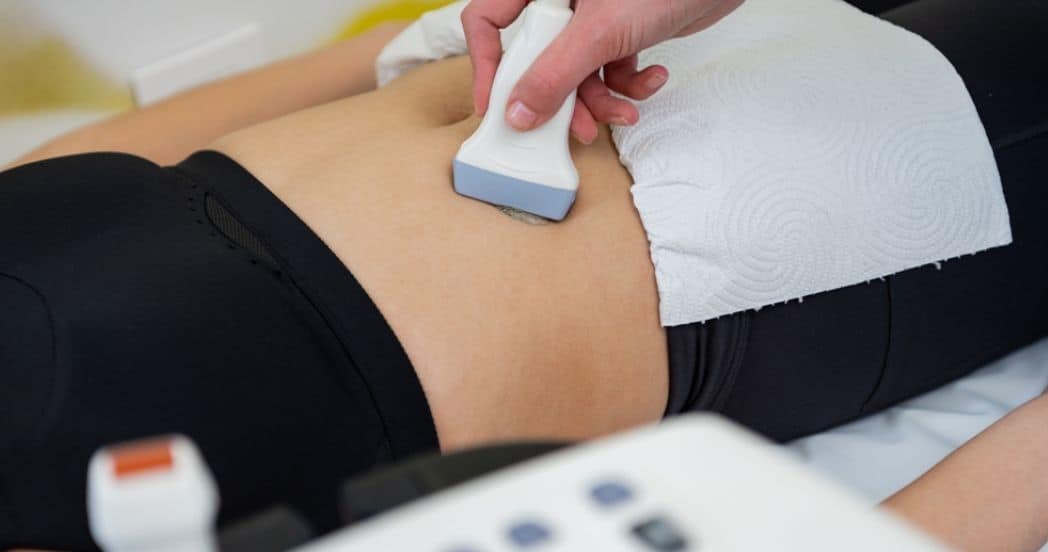
What Happens During an Antenatal Scan?
During ultrasound scan time, the procedure mainly includes:
Preparation:
You need to drink water beforehand to fill the bladder, improving the image’s clarity.
Procedure:
● You will be lying on an exam table where a gel is mainly applied to the abdomen.
● Next, a transducer is moved over the skin to find the images.
● When it comes to transvaginal ultrasound, the probe is gently inserted into the vagina for a significantly closer view.
Duration:
Generally, according to the purpose, most of the scans mainly take between 15 to 30 minutes.
Results:
The sonographer will discuss or forward the findings to the doctor for interpretation.
Benefits of Antenatal Scans
Reassurance: Witnessing your baby’s heart beating and movements could be an emotional and reassuring experience.
Early Detection: Finding potential problems at the earliest permits for timely interventions.
Monitoring Health: Frequent scans confirm that the baby and mother stay healthy throughout pregnancy.
Potential Side Effects of Ultrasound Scans
Ultrasound scans are considered to be safest and do not utilize radiation. However, some women could experience less discomfort during transvaginal ultrasound or transducer pressure. In this case, you can consult your healthcare provider for concerns.
What Can Be Detected Through Antenatal Scans?
Growth and Heartbeat: The baby’s size, weight and heart rate could be assessed.
Amniotic Fluid Levels: These are vital for the baby’s lung and movement development.
Placenta position: Placental location and health are vital for pregnancy.
Structural Problems: Abnormalcy in the spine, heart, and brain can be known.
How to Ready for Antenatal Scans
Ask Queries: Don’t delay to talk about issues with the doctor.
Wear Comfortable Clothing: Two piece outfits make it easy to approach the abdomen.
Check Instructions: Drink the suggested amount of water if needed.
Common Myths About Antenatal Scans
- Ultrasound Scans Are Harmful
Ultrasounds utilize sound waves, not radiation, and are mainly safe when it comes to babies and mothers.
- Scans Guarantee a Problem-Free Pregnancy
While scans offer valuable insights, they cannot predict all pregnancy outcomes.
- Drinking Much Water Is Important
Only monitor your healthcare provider’s advice related to hydration for the scan.
When to Schedule Antenatal Scans
Different weeks of pregnancy demand specific scans:
Early pregnancy: For confirmation, around 6 to 8 weeks.
Mid pregnancy: For anomaly scan, it will be around 18 to 22 weeks.
Late pregnancy: Monitor position and growth after 28 weeks.
Selecting the Right Facility
Choose a good clinic with skilled sonographers for antenatal scans. Confirm the facility delivers advanced imaging techniques and thorough prenatal evaluations.
Conclusion
Antenatal scans are a basis of prenatal care, offering invaluable insights into the development and unborn babies health. From deciding the due date with a dating scan to measuring blood flow with a respective transvaginal ultrasound, these scans confirm a baby, and you are on the right way towards a healthy and safe delivery.